



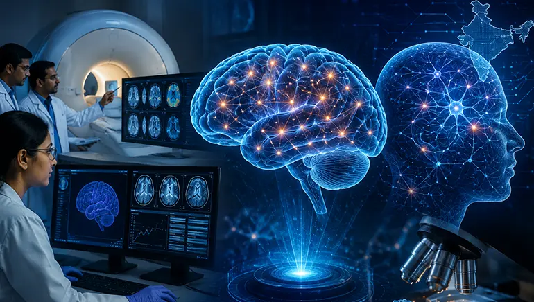
Indian scientists are playing a key role in addressing one of neuroscience’s biggest challenges, creating highly detailed maps of the human brain to understand better how it works and why neurological diseases develop.
Researchers are using cutting-edge imaging techniques alongside artificial intelligence (AI) to study brain tissue at an unprecedented level of detail. The effort is helping scientists trace millions of brain cells and their connections, offering new insights into diseases like Alzheimer’s and Parkinson’s, and disorders like epilepsy and autism.
Unlike conventional MRI scans that reveal only larger brain structures, the new approach captures microscopic details, allowing researchers to examine neurons, blood vessels and supporting cells in three dimensions. Scientists believe these high-resolution maps could transform the way brain diseases are diagnosed and treated.
The work is part of a broader international effort involving institutions globally to build comprehensive digital brain atlases. Researchers say the technology will help identify subtle changes that occur in the brain, potentially leading to earlier diagnosis and more targeted therapies.
Beyond medicine, the project is expected to deepen scientists’ understanding of how memory, learning, and cognition function. The data generated could also support future advances in AI, brain-computer interfaces and precision neuroscience.
Although mapping the human brain remains one of science’s most complex challenges, researchers believe each breakthrough brings them closer to unlocking the mysteries of the body’s most sophisticated organ. By combining advanced microscopy, computational analysis and AI, Indian scientists are helping shape the next generation of brain research.